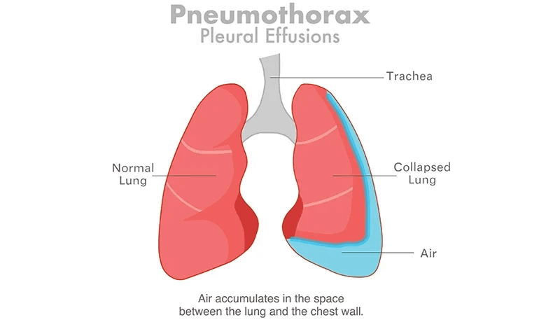
Pleural effusion is an abnormal buildup of fluid around the lung, in the narrow space between the lung and the inside of the chest wall.
It is sometimes described as “water on the lungs,” but that phrase can be misleading. The fluid usually collects outside the lung in the pleural space, rather than inside the air sacs.
At Jindal Chest, patients with unexplained breathlessness, chest pain or abnormal chest imaging may be evaluated for pleural conditions such as pleural effusion, infection, tuberculosis or other underlying lung diseases.
A small amount of pleural fluid is normally present and helps the lungs move smoothly during breathing. A pleural effusion develops when more fluid enters this space than the body can remove.
Some effusions are small and cause no symptoms. Others can become large enough to compress the lung and make breathing difficult.
Most importantly, pleural effusion is not a diagnosis by itself. It is a sign that another condition is affecting the lungs, pleura, heart or another part of the body.
Finding the cause is therefore a key part of treatment.
Pleural Effusion at a Glance
|
Question |
Quick answer |
|
What is pleural effusion? |
Excess fluid between the lung and chest wall |
|
Common symptoms |
Breathlessness, chest pain, dry cough |
|
Common causes |
Heart failure, pneumonia, tuberculosis, cancer, kidney or liver disease |
|
How is it diagnosed? |
Chest X-ray, ultrasound, CT and sometimes thoracentesis |
|
Does every effusion need drainage? |
No |
|
Can it return? |
Yes, depending on the cause |
|
Which specialist treats it? |
A pulmonologist or chest specialist |
What Is the Pleural Space?
Each lung is covered by a thin membrane called the visceral pleura.
Another membrane, called the parietal pleura, lines the inside of the chest wall.
The narrow area between these two layers is known as the pleural space.
Normally, it contains only a small amount of lubricating fluid.
When an abnormal amount of fluid accumulates in this space, the condition is called a pleural effusion.
Is Pleural Effusion the Same as Water in the Lungs?
Not exactly.
The phrase “water in the lungs” is used loosely for several different conditions.
In pleural effusion, fluid collects mainly outside the lung, between the pleural layers.
This is different from pulmonary oedema, where fluid accumulates within the lung tissue and air sacs.
The two conditions can occur together, particularly in conditions such as heart failure, but they are not the same problem.
What Are the Symptoms of Pleural Effusion?
Symptoms depend on:
· how much fluid has accumulated
· how quickly it developed
· whether one or both sides are affected
· the underlying cause
· the person's existing lung and heart health
Common symptoms include:
Shortness of Breath
Breathlessness is one of the most common symptoms.
A larger pleural effusion can compress the lung and limit how fully it expands.
Some people notice breathlessness only while walking or climbing stairs, while larger effusions may make breathing difficult even at rest.
Chest Pain
Pleural inflammation can cause pleuritic chest pain.
This is usually sharp and may become worse when:
· taking a deep breath
· coughing
· sneezing
In some cases, the pain may decrease as more fluid accumulates because the inflamed pleural surfaces are no longer rubbing directly against each other.
Dry Cough
Some patients develop a persistent dry cough.
Difficulty Breathing While Lying Down
This may occur particularly when the pleural effusion is related to heart failure.
Fever or Chills
Fever and chills may suggest infection such as pneumonia, pleural infection or tuberculosis.
However, fever is not present in every infectious effusion.
Unexplained Weight Loss or Poor Appetite
These symptoms may require investigation for conditions such as tuberculosis, cancer or other systemic illnesses.
Can Pleural Effusion Cause No Symptoms?
Yes.
A small pleural effusion may be discovered incidentally during a chest X-ray, ultrasound or CT scan performed for another reason.
However, the absence of symptoms does not automatically mean the effusion should be ignored.
A new or unexplained pleural effusion still needs to be interpreted in the context of the patient's medical history, imaging and likely cause.
What Causes Pleural Effusion?
Pleural effusion can result from many different conditions.
Some directly affect the lungs or pleura, while others originate in the heart, liver, kidneys or elsewhere in the body.
Common causes include:
· heart failure
· pneumonia
· tuberculosis
· cancer
· pulmonary embolism
· kidney disease
· liver cirrhosis
· autoimmune diseases
· pancreatitis
· chest trauma
· certain medications
Doctors often classify pleural fluid as either transudative or exudative because this helps narrow down the cause.
What Is a Transudative Pleural Effusion?
A transudative pleural effusion usually develops because of changes in pressure within blood vessels or low blood protein levels.
The pleura itself is not necessarily inflamed.
Common causes include:
Heart Failure
Heart failure is one of the most common causes of pleural effusion.
Fluid may accumulate on both sides of the chest, although this is not always the case.
Liver Cirrhosis
Advanced liver disease can cause fluid to move from the abdomen through the diaphragm into the pleural space.
This is known as hepatic hydrothorax.
Kidney Disease
Advanced kidney disease and conditions associated with very low blood protein can also contribute to pleural-fluid accumulation.
What Is an Exudative Pleural Effusion?
An exudative pleural effusion usually develops because the pleura is inflamed, injured or affected by abnormal cells.
Common causes include:
· pneumonia
· tuberculosis
· cancer
· pulmonary embolism
· autoimmune disease
· pancreatitis
· other infections
· certain medications
Exudative effusions usually require more investigation to identify the underlying cause.
Transudative vs Exudative Pleural Effusion
|
Feature |
Transudative |
Exudative |
|
Main mechanism |
Pressure or protein imbalance |
Inflammation, infection, malignancy or tissue injury |
|
Common causes |
Heart failure, cirrhosis, kidney disease |
Pneumonia, TB, cancer, pulmonary embolism |
|
Pleural inflammation |
Usually not the main problem |
Often present |
|
Need for further testing |
Diagnostic aspiration may not always be needed when the cause is clinically obvious and the effusion responds as expected to treatment |
Often requires fluid analysis and further investigation |
These categories help guide diagnosis, but they should never be interpreted in isolation.
Tuberculosis and Pleural Effusion
Tuberculosis is an important consideration in pleural effusion, particularly in India.
Pleural tuberculosis occurs when TB affects the membranes around the lung.
Possible symptoms include:
· fever
· chest pain
· cough
· breathlessness
· night sweats
· reduced appetite
· unexplained weight loss
Pleural TB usually produces an exudative effusion.
Evaluation may include:
· pleural fluid protein and LDH
· differential cell count
· adenosine deaminase, or ADA
· microbiological testing for tuberculosis
· cytology
· pleural biopsy in selected cases
An elevated ADA level can support a diagnosis of pleural TB when the clinical picture fits.
However, high ADA does not automatically prove tuberculosis.
Other diseases can also raise pleural-fluid ADA.
If the diagnosis remains unclear, pleural biopsy or thoracoscopy may be required.
Pneumonia and Pleural Effusion
Pneumonia can cause fluid to collect around the affected lung.
This is known as a parapneumonic effusion.
Some are small and improve when the pneumonia is treated.
Others become complicated or infected and may require drainage.
What Is Empyema?
Empyema means infected material or pus has been collected in the pleural space.
It is a serious form of pleural infection.
Possible features include:
· fever
· persistent illness despite antibiotics
· chest pain
· breathlessness
· pus in pleural fluid
· low pleural-fluid pH
· loculated or septated fluid on ultrasound or CT
Empyema generally requires drainage in addition to antibiotic treatment.
Antibiotics alone may not be sufficient when infected fluid remains trapped within the pleural space.
Pleural Effusion and Cancer
Several cancers can cause pleural effusion, including:
· breast cancer
· lymphoma
· cancers that spread to the pleura from other organs
A malignant pleural effusion occurs when cancer involves the pleura or malignant cells are identified in pleural fluid.
Pleural-fluid cytology may establish the diagnosis.
However, a negative cytology result does not always exclude malignancy.
If suspicion remains high, further evaluation may include:
· CT imaging
· repeat sampling in selected cases
· pleural biopsy
· thoracoscopy
Malignant pleural effusions can recur after simple drainage, so longer-term treatment may sometimes be required.
Can a Blood Clot Cause Pleural Effusion?
Yes.
A pulmonary embolism, which is a blood clot affecting the pulmonary circulation, can cause pleural effusion.
The effusion is often relatively small.
Possible accompanying symptoms include:
· sudden breathlessness
· chest pain
· rapid heartbeat
· coughing blood
· leg pain or swelling if deep-vein thrombosis is present
Suspected pulmonary embolism requires urgent medical assessment.
How Is Pleural Effusion Diagnosed?
Diagnosis usually begins with:
· a detailed medical history
· physical examination
· chest imaging
A doctor may ask about:
· fever
· cough
· weight loss
· tuberculosis exposure
· previous cancer
· heart disease
· liver or kidney disease
· current medicines
· blood-clot risk
· recent infection or surgery
The next step depends on the suspected cause.
What Can a Doctor Find on Examination?
A moderate or large pleural effusion may cause:
· reduced breath sounds
· dullness when the chest is tapped
· reduced movement of the affected side of the chest
Physical examination can suggest an effusion, but it cannot reliably determine the cause.
Imaging is usually necessary.
Chest X-Ray for Pleural Effusion
A chest X-ray is often the first imaging test.
It can show:
· fluid collecting at the bottom of the chest
· blunting of the costophrenic angle
· compression of the lung in larger effusions
· associated lung abnormalities
Very small effusions may not be obvious on a standard chest X-ray.
Ultrasound for Pleural Effusion
Chest ultrasound is particularly useful because it can:
· detect relatively small amounts of fluid
· estimate the size of the effusion
· identify septations or loculations
· distinguish fluid from some solid abnormalities
· help identify a safer site for thoracentesis
Ultrasound guidance is preferred for pleural aspiration because it reduces procedure-related complications.
CT Scan for Pleural Effusion
A CT scan provides more detailed information about:
· lung tissue
· pleural thickening
· masses
· lymph nodes
· loculated fluid
· pneumonia
· possible malignancy
· other causes of breathlessness
CT is especially useful when the cause remains uncertain or when malignancy, complicated infection or another pleural disorder is suspected.
What Is Thoracentesis?
Thoracentesis, also called pleural aspiration or pleurocentesis, is a procedure in which a needle or catheter is introduced through the chest wall into the pleural space.
It may be performed for two different reasons.
Diagnostic Thoracentesis
A sample of fluid is removed and sent for testing to help determine the cause.
Therapeutic Thoracentesis
A larger amount of fluid may be removed to relieve symptoms such as significant breathlessness.
Thoracentesis should generally be performed with ultrasound guidance when appropriate.
Does Every Pleural Effusion Need Thoracentesis?
No.
This is an important distinction.
For example, a patient with a small bilateral pleural effusion and clinically obvious heart failure that improves as expected with treatment may not always require diagnostic aspiration.
Thoracentesis is more likely to be considered when an effusion is:
· new
· unexplained
· mainly on one side
· more than minimal
· associated with pneumonia
· associated with suspected cancer
· associated with suspected tuberculosis
· not responding as expected to treatment
The decision should always be individualised.
What Tests Are Done on Pleural Fluid?
Depending on the clinical situation, pleural fluid may be tested for:
· protein
· lactate dehydrogenase, or LDH
· glucose
· pH
· cell count
· Gram stain
· bacterial culture
· cytology
· TB-related investigations
· ADA
· triglycerides
· other specialised tests
Blood tests are often performed at the same time so serum and pleural-fluid values can be compared.
What Are Light's Criteria?
Light's criteria help determine whether pleural fluid is exudative.
An effusion is classified as exudative if at least one of the following is present:
1. Pleural-fluid protein / serum protein ratio greater than 0.5
2. Pleural-fluid LDH / serum LDH ratio greater than 0.6
3. Pleural-fluid LDH greater than two-thirds of the upper limit of normal serum LDH
These criteria help guide the diagnostic pathway.
Patients do not need to calculate the values themselves. They should be interpreted along with the medical history, imaging and other test results.
Why Does Pleural-Fluid pH Matter?
Pleural-fluid pH is particularly important when pneumonia or pleural infection is suspected.
When the fluid is not obviously pus, a pH of 7.2 or lower suggests a higher risk of complicated parapneumonic effusion or pleural infection.
When there is enough safely accessible fluid, chest-tube drainage is generally considered in this situation.
Other findings such as persistent fever, low glucose, high LDH and septations on ultrasound also influence treatment decisions.
When Is a Chest Tube Needed?
A chest tube, also called an intercostal drain, may be required when:
· pus is present
· pleural infection is established
· a complicated parapneumonic effusion requires drainage
· blood has collected in the pleural space
· a large or recurrent collection cannot be managed adequately with simple aspiration
· another specific clinical indication exists
The decision depends on the cause and clinical situation, not simply on the volume of fluid.
When Is Thoracoscopy Needed?
Not every pleural effusion can be diagnosed through imaging and fluid analysis alone.
Thoracoscopy allows a pulmonologist to directly inspect the pleural cavity using an endoscope.
Abnormal areas of the pleura can be biopsied under direct vision.
Thoracoscopy may be particularly useful when:
· an unexplained exudative effusion persists
· cancer is suspected
· tuberculosis remains possible despite inconclusive fluid tests
· pleural thickening or nodules are present
· a tissue diagnosis is required
At Jindal Chest, thoracoscopy may be considered in selected patients when pleural fluid analysis and imaging do not establish the cause of an unexplained pleural effusion or when pleural tissue is required for diagnosis.
Thoracoscopy may also be used for selected therapeutic procedures.
How Is Pleural Effusion Treated?
There is no single treatment for pleural effusion.
Treatment depends on why the fluid accumulated.
Heart-Failure-Related Pleural Effusion
Treatment generally focuses on controlling the underlying heart failure.
This may include diuretic therapy and other cardiovascular treatment.
Large symptomatic effusions may sometimes require drainage.
Pneumonia-Related Pleural Effusion
Treatment may include:
· antibiotics
· monitoring
· pleural-fluid sampling
· chest-tube drainage if the fluid becomes complicated or infected
Tuberculous Pleural Effusion
Confirmed pleural tuberculosis requires appropriate anti-tuberculosis treatment.
Drainage may be required in selected situations, particularly when the collection is large, symptomatic or complicated.
Malignant Pleural Effusion
Treatment depends on:
· the underlying cancer
· symptoms
· whether the fluid returns
· how well the lung re-expands
· the patient's overall condition
Options may include:
· drainage
· indwelling pleural catheter
· pleurodesis
· cancer-directed treatment
· other pleural procedures
Kidney or Liver Disease
Treatment mainly focuses on managing the underlying systemic condition.
Pulmonary Embolism
Treatment targets the blood clot and its underlying risk factors.
Small associated pleural effusions do not automatically require drainage.
What Is Pleurodesis?
Pleurodesis is a procedure intended to prevent fluid or air from repeatedly accumulating in the pleural space.
A substance is introduced into the pleural cavity to encourage the pleural surfaces to adhere to each other.
It may be considered in selected patients with recurrent effusions, particularly certain malignant pleural effusions.
Whether pleurodesis is appropriate depends on:
· the underlying disease
· symptom burden
· how quickly fluid returns
· whether the lung re-expands after drainage
Can Pleural Effusion Come Back?
Yes.
Whether it returns depends mainly on the underlying cause.
For example:
· heart-failure effusions may recur if heart failure remains poorly controlled
· malignant pleural effusions can recur unless longer-term management is used
· infection-related effusions may resolve after adequate treatment
· TB-related effusions require appropriate tuberculosis treatment
· recurrent unexplained effusions require further investigation
Repeatedly draining pleural fluid without identifying and treating the cause is usually not an adequate long-term strategy.
Is Pleural Effusion Dangerous?
It can be.
The seriousness depends on:
· the underlying cause
· the amount of fluid
· how quickly it accumulated
· whether infection is present
· its effect on breathing
· the patient's other medical conditions
For example, a small effusion caused by controlled heart failure is very different from an infected empyema.
Pleural effusion should therefore never be judged by size alone.
Can Pleural Effusion Be Cancer?
Yes, but not every pleural effusion is caused by cancer.
Other common causes include:
· heart failure
· pneumonia
· tuberculosis
· kidney disease
· liver disease
· pulmonary embolism
Cancer becomes more concerning when an effusion is associated with:
· unexplained recurrence
· pleural thickening or nodularity
· unexplained weight loss
· known cancer
· suspicious CT findings
Even then, a tissue or fluid diagnosis may be needed before conclusions are drawn.
Can Pleural Effusion Be Caused by Tuberculosis?
Yes.
Pleural TB is an important cause of exudative pleural effusion, particularly in regions where tuberculosis remains common.
Diagnosis may require more than a chest X-ray.
Pleural-fluid analysis, TB testing and sometimes pleural biopsy or thoracoscopy may be required.
Can Pleural Effusion Resolve Without Drainage?
Yes, in some cases.
For example, small effusions caused by heart failure may improve when the heart condition is treated.
Some small uncomplicated parapneumonic effusions may resolve with appropriate antibiotics.
Drainage is more likely to be required when:
· the effusion causes significant breathlessness
· infection is complicated
· pus is present
· diagnostic fluid is needed
· fluid repeatedly returns
· another condition requires direct pleural treatment
Can Pleural Effusion Be Prevented?
There is no single method that prevents every pleural effusion because the causes are so varied.
Risk may sometimes be reduced through appropriate management of:
· heart failure
· kidney disease
· liver disease
· pneumonia
· tuberculosis
Avoiding tobacco exposure and seeking evaluation for persistent respiratory symptoms can also support overall lung health.
When Should You Seek Urgent Medical Care?
Seek urgent medical attention for:
· severe or rapidly worsening breathlessness
· sudden unexplained chest pain
· blue or grey lips
· fainting
· severe weakness
· coughing blood
· high fever with breathing difficulty
· very low oxygen levels
· symptoms suggesting pulmonary embolism
These symptoms may indicate a serious pleural condition or another medical emergency.
When Should You See a Pulmonologist?
A pulmonologist's assessment is particularly useful when:
· an effusion is unexplained
· fluid repeatedly returns
· tuberculosis is suspected
· pneumonia is not improving as expected
· cancer is being considered
· thoracentesis is required
· pleural biopsy may be required
· thoracoscopy is being considered
The goal is not simply to remove the fluid.
The more important task is to determine why the fluid accumulated and whether the underlying disease needs specific treatment.
If you have been diagnosed with an unexplained or recurrent pleural effusion, or your chest X-ray or CT shows fluid around the lung, you can consult the pulmonology team at Jindal Chest for further evaluation.
Frequently Asked Questions
What is pleural effusion in simple terms?
Pleural effusion is an abnormal collection of fluid in the space between the lung and chest wall.
What is the most common cause of pleural effusion?
There is no single cause in every patient.
Heart failure is an important cause of transudative effusions, while pneumonia, tuberculosis, malignancy and pulmonary embolism are important causes of exudative effusions.
Is pleural effusion the same as water in the lungs?
Not exactly.
With pleural effusion, fluid collects around the lung. In pulmonary oedema, fluid accumulates inside the lung tissue and air sacs.
Can pleural effusion be cured?
Many pleural effusions resolve when the underlying cause is successfully treated.
Whether it can be permanently resolved depends on why the fluid developed.
Does pleural effusion always need drainage?
No.
Small effusions with an obvious, treatable cause may improve without direct drainage.
Drainage may be needed for diagnosis, symptom relief or treatment of infection.
Is thoracentesis painful?
Local anaesthetic is normally used to numb the area.
Patients may feel pressure or temporary discomfort, but significant pain is not expected.
Is thoracentesis safe?
Thoracentesis is commonly performed and is generally safe when appropriately indicated.
Possible complications include pneumothorax, bleeding and infection.
Ultrasound guidance helps reduce procedure-related risk.
Is pleural effusion always tuberculosis?
No.
Tuberculosis is an important cause, particularly in India, but heart failure, pneumonia, cancer, kidney or liver disease and pulmonary embolism can also cause pleural effusion.
Does a high ADA level always mean tuberculosis?
No.
An elevated pleural-fluid ADA can support a diagnosis of pleural TB when the overall clinical picture fits, but it should not be interpreted alone.
Is pleural effusion always cancer?
No.
Cancer is one possible cause, but many pleural effusions are caused by non-cancerous conditions.
Imaging, fluid analysis and sometimes pleural biopsy may be needed when malignancy is suspected.
What is the difference between pleural effusion and pneumonia?
Pneumonia is an infection of the lung tissue.
Pleural effusion is fluid around the lung.
Pneumonia can itself cause a pleural effusion, known as a parapneumonic effusion.
What is the difference between pleural effusion and empyema?
Pleural effusion is the general term for abnormal excess fluid around the lung.
Empyema refers specifically to infected pleural fluid or pus and generally requires drainage along with antibiotics.
Can pleural effusion return after thoracentesis?
Yes.
Thoracentesis removes fluid but does not necessarily eliminate the underlying cause.
If that cause remains active, the fluid can return.
Is thoracoscopy required for every pleural effusion?
No.
Thoracoscopy is generally reserved for selected cases where imaging and fluid analysis do not provide a diagnosis or where pleural tissue is required.
Conclusion
Pleural effusion is an abnormal accumulation of fluid around the lung, but the fluid itself is only part of the problem.
The key question is why it accumulated.
Common causes include:
· heart failure
· pneumonia
· tuberculosis
· cancer
· kidney or liver disease
· pulmonary embolism
Chest X-ray, ultrasound and sometimes CT help identify and assess the effusion. When necessary, ultrasound-guided thoracentesis allows doctors to analyse the fluid and can also relieve significant breathlessness.
Some effusions improve when the underlying condition is treated. Others require drainage, a chest tube, pleural biopsy, thoracoscopy or longer-term treatment to reduce recurrence.
At Jindal Chest, the focus in patients with pleural effusion is not simply on removing the fluid, but on identifying and treating the condition responsible for it. Depending on the clinical situation, evaluation may include chest imaging, ultrasound-guided pleural aspiration, fluid analysis, thoracoscopy or other respiratory investigations.
A new, unexplained or recurrent pleural effusion should therefore be properly evaluated rather than repeatedly drained without establishing the underlying cause.
