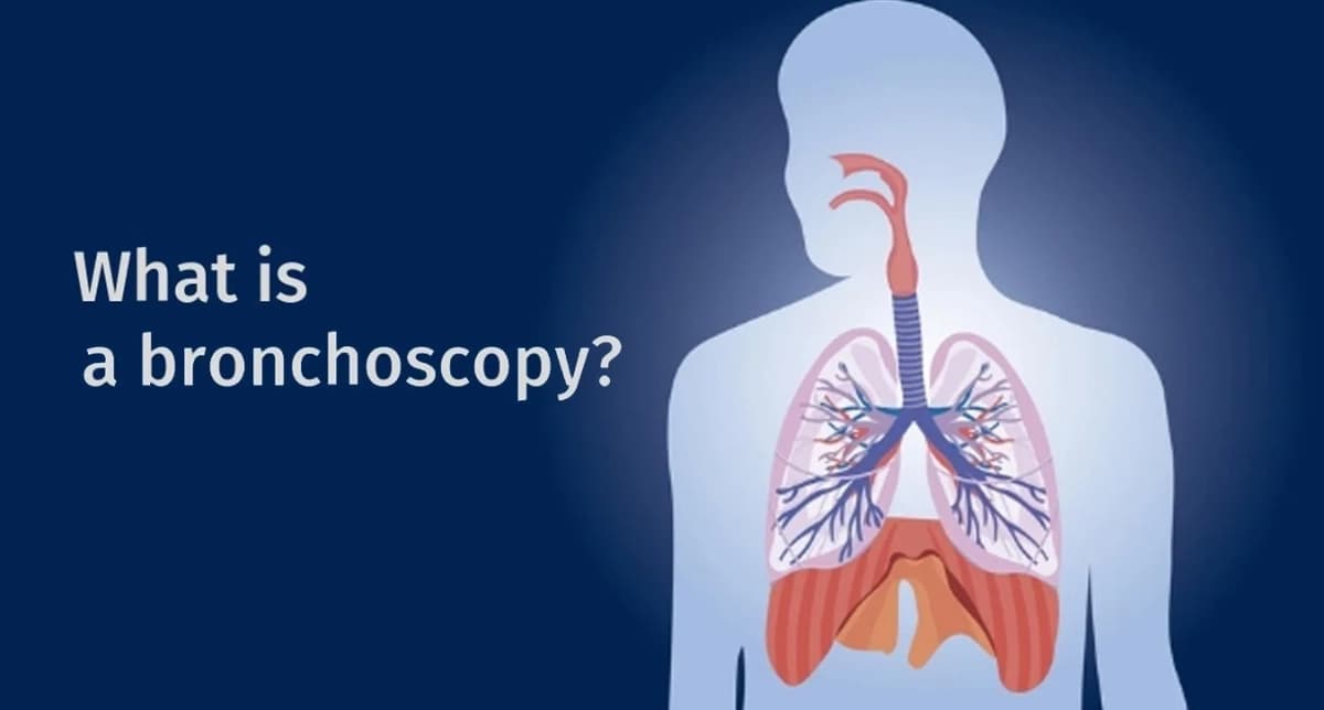
What Is Bronchoscopy? Procedure, Uses, Risks and Recovery
Bronchoscopy is a procedure in which a lung specialist passes a thin, flexible tube with a camera through the nose or mouth and into the airways. It allows the doctor to examine the windpipe and bronchi, collect fluid or tissue samples, and sometimes remove mucus plugs, foreign bodies or other airway obstructions. Flexible bronchoscopy is usually performed with local anaesthesia, with or without sedation, and many patients can go home the same day.
Being advised to have a bronchoscopy can sound worrying, especially when words such as biopsy, tuberculosis or lung cancer have also been mentioned.
In practice, flexible bronchoscopy is a commonly used respiratory procedure. What you experience, how long it takes and its risks depend mainly on why the bronchoscopy is being performed and which samples or treatments are required.
A large study from the Department of Pulmonary Medicine at PGIMER, Chandigarh reviewed 9,979 flexible bronchoscopies performed under local anaesthesia without sedation between 2013 and 2021. Major complications occurred in fewer than 1% of procedures.
This guide explains why bronchoscopy is performed, what happens during the procedure, how samples such as BAL and biopsies are obtained, possible risks and what recovery is usually like.
Why would a doctor recommend bronchoscopy?
A doctor may recommend bronchoscopy when symptoms, a chest X-ray or a CT scan suggest that the airways or lungs need to be examined directly or sampled.
Common reasons include:
-
An abnormal shadow, mass or other finding on a chest X-ray or CT scan
-
Suspected lung cancer
-
Suspected tuberculosis when appropriate sputum testing has not provided an answer
-
Pneumonia or another lung infection that is not resolving as expected
-
Coughing up blood
-
Enlarged lymph nodes inside the chest
-
Suspected sarcoidosis
-
A collapsed part of the lung caused by an airway blockage
-
Removing thick secretions or mucus plugs
-
Removing an inhaled foreign body
-
Investigating selected unexplained respiratory symptoms
In the large PGIMER study, the leading indications included suspected lung cancer, tuberculosis and sarcoidosis. However, those figures describe patients at a major tertiary referral centre in Chandigarh and should not be interpreted as the proportion of bronchoscopies performed for these diseases everywhere.
You can also read our overview of tests used to diagnose lung diseases to understand where bronchoscopy fits alongside chest X-rays, CT scans and lung-function tests.
What can doctors do during bronchoscopy?
Bronchoscopy is not simply a camera examination.
Small instruments can be passed through the bronchoscope to collect different types of samples. The technique chosen depends on what the doctor is trying to diagnose.
|
Procedure |
What happens |
Common uses |
|
Airway inspection |
The doctor directly examines the trachea and bronchi |
Bleeding, suspected obstruction, airway abnormalities |
|
Bronchoalveolar lavage (BAL) |
Sterile saline is placed into part of the lung and suctioned back for testing |
Tuberculosis, fungal or other infections and selected inflammatory lung diseases |
|
Endobronchial biopsy (EBB) |
Small tissue samples are taken from an abnormality visible inside the airway |
Lung cancer, endobronchial TB, sarcoidosis and other lesions |
|
Transbronchial lung biopsy (TBLB/TBB) |
Small samples are obtained from lung tissue beyond the visible bronchial wall |
Sarcoidosis, selected interstitial lung diseases and infections |
|
TBNA |
A needle is passed through the airway wall to obtain samples |
Selected lymph nodes or masses |
|
EBUS-TBNA |
Ultrasound at the tip of a specialised bronchoscope guides needle sampling in real time |
Lung-cancer staging, tuberculosis, sarcoidosis and other enlarged lymph-node conditions |
|
Therapeutic bronchoscopy |
Instruments are used to remove material or treat an airway problem |
Foreign bodies, mucus plugs, bleeding or selected airway obstruction |
For more information about ultrasound-guided lymph-node sampling, see our page on endobronchial ultrasound, or EBUS.
Flexible vs rigid bronchoscopy
Most diagnostic bronchoscopies are performed using a flexible bronchoscope.
It is a narrow, manoeuvrable instrument that can pass through the nose or mouth and reach different branches of the airway.
A rigid bronchoscope is a straight hollow instrument generally used under general anaesthesia. It may be needed for situations such as:
-
Removal of certain large or difficult foreign bodies
-
Control of significant airway bleeding
-
Treatment of major central-airway obstruction
-
Placement of some airway stents
-
Other specialised interventional procedures
Which method is appropriate depends on the patient's condition and what needs to be done.
Can bronchoscopy remove something stuck in the lung?
Yes.
Bronchoscopy can be both diagnostic and therapeutic.
One example from Jindal Chest Clinics involved a 55-year-old man who developed persistent cough and an unusual pricking sensation after consuming bone soup. Imaging suggested an inhaled foreign body, and bronchoscopy found a piece of bone lodged in the right main bronchus.
It was removed bronchoscopically.
Foreign-body aspiration is more common in children but can also occur in adults, particularly when swallowing is impaired or consciousness is reduced.
How should you prepare for a bronchoscopy?
Preparation depends on the type of bronchoscopy, whether a biopsy is planned and whether sedation or general anaesthesia will be used.
Your treating team should give you specific instructions.
Tell your doctor about your medicines
Inform the doctor about all prescription medicines and supplements you take, particularly:
-
Aspirin
-
Clopidogrel
-
Warfarin
-
Apixaban
-
Rivaroxaban
-
Dabigatran
-
Other blood-thinning medicines
-
Diabetes medicines or insulin
Also mention any bleeding disorder, previous reaction to anaesthesia, drug allergy, heart disease or significant breathing problem.
Do not stop blood-thinning medication on your own.
Whether a medicine needs to be withheld depends on the procedure being planned and your reason for taking it.
Do you need to fast before bronchoscopy?
Yes, fasting is required to reduce the risk of stomach contents entering the lungs during the procedure.
Joint Indian bronchoscopy guidelines recommend a minimum fasting period of approximately:
-
2 hours for clear liquids
-
4 hours for a light meal
Longer fasting may be required for heavier meals, sedation, general anaesthesia or according to the hospital's protocol.
Therefore, follow the instructions given by your own bronchoscopy unit rather than using a general internet fasting rule.
Bring your reports
Bring relevant:
-
Chest X-rays
-
CT scans
-
Previous pathology reports
-
Blood-test reports
-
Current medication list
-
Previous respiratory records
If sedation is planned, arrange transport home according to the instructions provided by the clinic.
How is bronchoscopy performed?
The exact process varies between hospitals, but a routine flexible bronchoscopy commonly follows these steps.
1. Monitoring is attached
Your blood pressure, heart rate and blood oxygen level are monitored.
An intravenous line is usually placed.
2. Local anaesthetic is given
A local anaesthetic such as lignocaine is used to numb the nose, throat and airways and reduce coughing and discomfort.
If the bronchoscope is passed through the nose, anaesthetic gel may also be applied inside the nostril.
3. Sedation may be given
Some bronchoscopies are performed with local anaesthesia alone.
Others use light or moderate intravenous sedation to improve comfort and tolerance. More complex procedures may require deeper sedation or general anaesthesia.
The approach depends on the procedure, the patient's health, the facility and the bronchoscopist.
4. The bronchoscope enters the airway
The flexible bronchoscope is passed through the nose or mouth, past the vocal cords and into the trachea and bronchi.
You can continue breathing around the scope.
Coughing, pressure or an unusual sensation in the throat can occur, especially while the bronchoscope is being positioned.
5. The airways are examined and samples taken
The doctor examines the airways and performs the planned sampling procedure, such as BAL, biopsy or needle aspiration.
Tissue sampling is generally tolerated under appropriate local anaesthesia and sedation, although sensations vary between patients.
Tell the medical team immediately if you experience significant pain or breathing difficulty.
6. The bronchoscope is removed
Once examination and sampling are complete, the scope is withdrawn.
How long does bronchoscopy take?
A straightforward flexible bronchoscopy may take approximately 15 to 30 minutes, although the time varies.
The bronchoscopy service at Jindal Chest Clinics describes routine procedures taking around 15 to 20 minutes.
The procedure can take longer when:
-
Several types of samples are required
-
EBUS-TBNA is performed
-
A foreign body needs to be removed
-
Bleeding needs to be controlled
-
Another therapeutic intervention is required
Preparation and recovery mean that your total time at the clinic or hospital will be longer than the bronchoscopy itself.
You can read more about the procedure on our bronchoscopy service page.
Will I be awake during bronchoscopy?
Possibly.
Flexible bronchoscopy may be performed with:
-
Local anaesthesia alone
-
Local anaesthesia with conscious or moderate sedation
-
Deep sedation in selected cases
-
General anaesthesia for certain procedures
Practice varies between centres and according to the procedure.
The large PGIMER study cited in this article specifically examined bronchoscopies performed without intravenous sedation, so those results should not be assumed to represent every bronchoscopy setting.
Ask your doctor which type of anaesthesia or sedation is planned for you.
Is bronchoscopy painful?
Bronchoscopy is generally described as uncomfortable rather than painful.
Common sensations include:
-
Numbness in the nose or throat
-
An unusual taste from the local anaesthetic
-
An urge to cough
-
Gagging
-
Pressure or awareness of the scope
-
Temporary throat irritation afterwards
Local anaesthesia and, when appropriate, sedation are used to improve comfort.
Individual experiences differ, particularly when different sampling techniques or therapeutic procedures are performed.
What are the risks of bronchoscopy?
Flexible bronchoscopy is generally safe, but it is an invasive procedure and complications can occur.
The risk depends substantially on what is done through the bronchoscope.
Possible complications include:
-
Bleeding
-
Temporary fall in blood oxygen
-
Pneumothorax, or air leaking around the lung
-
Bronchospasm
-
Abnormal heart rhythm
-
Reactions to sedation or medications
-
Fever after bronchoscopy
-
Infection, although significant procedure-related infection is uncommon
-
Rare serious cardiopulmonary complications
What does the PGIMER study show?
Researchers at PGIMER reviewed 9,979 flexible bronchoscopies performed under local anaesthesia without intravenous sedation.
Major complications were uncommon.
|
Outcome |
Reported frequency |
|
Any major complication |
Approximately 0.93% |
|
Pneumothorax |
0.44% |
|
Severe bleeding |
0.27% |
|
Endotracheal intubation |
0.23% |
|
Death |
0.08% |
Among the 44 patients who developed pneumothorax, 11 required insertion of a chest drain.
The risk also increased with more invasive sampling.
Airway inspection alone was associated with considerably fewer complications than procedures involving transbronchial lung biopsy.
Importantly, these figures came from a tertiary referral hospital treating many patients with serious respiratory illnesses. The researchers themselves cautioned that the results may not be generalisable to every hospital or individual patient.
They should therefore be used to understand the overall safety profile of bronchoscopy, not to calculate an individual patient's personal risk.
Which type of bronchoscopy carries greater risk?
An inspection-only bronchoscopy generally carries less risk than a procedure involving lung tissue sampling.
In the PGIMER study, transbronchial biopsy was particularly associated with a higher risk of major complications compared with inspection alone.
That does not mean a biopsy should be avoided when it is needed.
The relevant question is whether obtaining tissue is likely to provide information that changes diagnosis or treatment.
Before the procedure, you can ask:
-
What sample are you planning to take?
-
Why is that sample necessary?
-
What is the main risk of this particular sampling method?
-
Are there alternatives?
-
How will the result affect my treatment?
What happens after bronchoscopy?
You are usually monitored until the medical team is satisfied that you are stable and any sedative medication has sufficiently worn off.
Because local anesthetic temporarily reduces the normal sensation and protective reflexes in your throat, do not eat or drink until your medical team confirms that it is safe to swallow.
Many centres wait until throat sensation and the cough or gag reflex have returned, often around one to two hours.
Once swallowing is safe, you may usually start with sips of water.
What is normal after bronchoscopy?
Temporary symptoms can include:
-
Sore throat
-
Hoarse voice
-
Mild cough
-
Small streaks of blood in sputum after some biopsies
-
Temporary tiredness after sedation
-
Mild transient fever in some patients
These usually improve.
Your treating team should explain what to expect based on the samples taken during your particular procedure.
Do you need a chest X-ray after a bronchoscopy?
Not everyone does.
A chest X-ray may be obtained if there is concern about pneumothorax, particularly after procedures that sample peripheral lung tissue or if symptoms develop.
Routine post-procedure imaging policies vary between units and procedures.
New chest pain, increasing breathlessness or a fall in oxygen after bronchoscopy requires prompt assessment.
When should you seek urgent medical attention after bronchoscopy?
Contact your treating team or seek urgent medical care if you develop:
-
Increasing or significant coughing of fresh blood
-
New or worsening breathlessness
-
Significant or increasing chest pain
-
Persistent fever
-
Fainting or severe weakness
-
Persistent vomiting
-
Unusual or prolonged drowsiness after sedation
-
Any symptom your bronchoscopy team specifically told you to watch for
Minor blood-streaking after certain biopsies is different from substantial or increasing bleeding.
If you are unsure whether the amount of bleeding is expected, contact the treating team rather than waiting.
Can you drive after bronchoscopy?
If you received sedation, you should not drive yourself home.
Restrictions after sedation commonly include avoiding driving, alcohol, operating machinery and important legal decisions for approximately 24 hours, although you should follow the exact instructions supplied by your hospital.
If no sedative medicine was used, restrictions may be different.
Do not assume that advice given to another patient applies to your procedure.
When do bronchoscopy results come?
Different samples have different processing times.
Some microbiological tests may produce early results within a few days, while:
-
Histopathology from a biopsy may take several days
-
Special stains or molecular tests can require additional time
-
Mycobacterial or fungal cultures may take several weeks
The exact turnaround time depends on the test and laboratory.
A bronchoscopy can still be useful even when the airways look normal because important information may come from lavage, biopsy or needle samples examined in the laboratory.
Can bronchoscopy diagnose tuberculosis?
Bronchoscopy can help diagnose pulmonary tuberculosis in selected patients, particularly when sputum cannot be obtained or when appropriate sputum investigations have not provided a diagnosis despite continuing clinical suspicion.
Samples such as bronchial wash or BAL may be tested using:
-
Smear microscopy
-
Molecular tests
-
Mycobacterial culture
-
Other microbiological methods
Bronchoscopy is not automatically required for every person being evaluated for tuberculosis.
The appropriate test sequence depends on symptoms, imaging and previous sputum investigations.
Can bronchoscopy diagnose lung cancer?
Yes, bronchoscopy is an important diagnostic tool for selected suspected lung cancers.
Depending on where the abnormality is located, doctors may obtain:
-
Endobronchial biopsy
-
Bronchial washings
-
Brush samples
-
Transbronchial biopsy
-
Conventional TBNA
-
EBUS-guided needle samples
These samples can help establish whether cancer is present and identify its pathological type.
Adequate tissue may also be used for molecular and biomarker testing when appropriate, which can influence treatment choices.
If enlarged lymph nodes need to be sampled for diagnosis or staging, EBUS may be particularly useful.
Is bronchoscopy surgery?
Flexible bronchoscopy is generally classified as an endoscopic procedure rather than open surgery.
The bronchoscope enters through the nose or mouth, so routine flexible bronchoscopy does not require a surgical cut in the chest.
However, that does not mean it is a trivial test.
It remains an invasive medical procedure requiring appropriate preparation, monitoring, consent and trained personnel.
Bronchoscopy mein dard hota hai kya?
Aam taur par flexible bronchoscopy ko log dard se zyada uncomfortable describe karte hain.
Naak, gale aur airways ko local anaesthetic se sunn kiya jata hai. Procedure ke dauraan khansi, gagging ya pressure jaisa feel ho sakta hai. Kuch patients ko sedation bhi di ja sakti hai.
Procedure kitni der chalega aur sedation ki zarurat hogi ya nahi, yeh is baat par depend karta hai ki sirf airways dekhni hain ya BAL, biopsy, EBUS ya koi treatment bhi karna hai.
Apne doctor se procedure se pehle poochhein ki aapke case mein exactly kya plan hai.
What questions should I ask before bronchoscopy?
Before agreeing to the procedure, useful questions include:
-
Why do I need a bronchoscopy?
-
What disease are you trying to confirm or rule out?
-
Which samples will you take?
-
Will I receive sedation?
-
How long should I fast?
-
Should I change any blood thinner or diabetes medicine?
-
What are the important risks in my case?
-
Will I need someone to take me home?
-
When should I expect the results?
-
What symptoms after the procedure should make me contact the clinic?
These answers are more useful than thinking of every bronchoscopy as the same procedure.
A short procedure can answer an important diagnostic question
A CT scan may show an abnormality, but imaging alone cannot always determine exactly what it represents.
Depending on the case, an abnormality may represent infection, inflammation, cancer or another condition requiring very different treatment.
Bronchoscopy allows pulmonologists to inspect the airways and, importantly, obtain appropriate samples for laboratory diagnosis.
If your doctor has recommended bronchoscopy, ask why it is needed, what sampling procedure is planned, what anaesthesia or sedation will be used, and when the results are expected.
For information about evaluation of persistent respiratory symptoms before invasive tests are considered, see our guide on when to consult a pulmonologist for persistent cough or breathlessness.
Jindal Chest Clinics, Sector 20D, Chandigarh, provides bronchoscopy and other advanced pulmonary diagnostic procedures.
Book an appointment or call 0172-4911000 or +91 9779030507.
