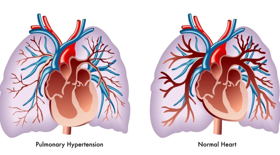
Pulmonary Hypertension: Symptoms, Causes and Treatment
Pulmonary hypertension means abnormally high pressure in the circulation between the right side of the heart and the lungs. It is defined by a mean pulmonary artery pressure above 20 mmHg at rest when measured directly by right-heart catheterisation. Common symptoms include increasing breathlessness during activity, fatigue, reduced exercise capacity, dizziness, chest discomfort and, in more advanced disease, fainting or swelling of the legs. Pulmonary hypertension has several different causes, so treatment depends on identifying the correct type rather than treating an echocardiogram number alone.
Pulmonary hypertension, or PH, is different from ordinary high blood pressure.
A blood-pressure cuff measures pressure in the arteries supplying the body. Pulmonary hypertension affects the circulation carrying blood from the right side of the heart through the lungs.
You can therefore have normal blood pressure in your arm and still have pulmonary hypertension.
Another important distinction is that pulmonary hypertension is not one disease.
It can result from left-sided heart disease, lung disease, chronic blood clots, pulmonary arterial hypertension or several less common conditions. Finding the cause is essential because treatments that help one type may be ineffective or potentially harmful in another.
What exactly is pulmonary hypertension?
Pulmonary hypertension is defined haemodynamically as a mean pulmonary artery pressure, or mPAP, above 20 mmHg at rest, measured during right-heart catheterisation.
That definition should not be confused with an estimated PASP or RVSP reported on an echocardiogram.
For pre-capillary pulmonary hypertension, additional measurements include:
-
Pulmonary arterial wedge pressure of 15 mmHg or less
-
Pulmonary vascular resistance above 2 Wood units
These measurements help doctors distinguish pulmonary arterial disease from pressure caused by the left side of the heart.
What are the symptoms of pulmonary hypertension?
Symptoms can be subtle at first.
The most common early problem is gradually increasing breathlessness during physical activity.
Other possible symptoms include:
-
Reduced exercise capacity
-
Unusual fatigue
-
Chest discomfort
-
Palpitations
-
Light-headedness
-
Dizziness during activity
-
Fainting or near-fainting
-
Swelling of the ankles or legs
-
Abdominal swelling in advanced right-heart failure
-
Breathlessness at rest in advanced disease
These symptoms are not specific to pulmonary hypertension.
Asthma, COPD, pulmonary fibrosis, anaemia, heart disease and physical deconditioning can produce similar symptoms.
Our guide to the common causes of shortness of breath explains why breathlessness needs a broader evaluation.
Why does pulmonary hypertension cause breathlessness?
The right side of the heart normally pumps blood into a low-pressure circulation through the lungs.
When resistance in that circulation rises, the right ventricle has to work harder.
During exercise, the heart may eventually become unable to increase blood flow enough to meet the body's demands. This contributes to:
-
Breathlessness
-
Fatigue
-
Reduced exercise capacity
-
Dizziness
If the condition becomes severe, the right ventricle can enlarge and weaken.
Fluid may then accumulate in the legs or abdomen.
Is fainting a warning sign?
Yes.
Fainting or near-fainting during exertion can occur in significant pulmonary hypertension and deserves prompt medical evaluation.
It is particularly concerning when accompanied by:
-
Chest pain
-
Rapidly increasing breathlessness
-
Blue or grey lips
-
Marked weakness
-
New swelling
-
Palpitations
Actual fainting, severe chest pain or severe breathlessness should be treated as urgent symptoms rather than waiting for a routine appointment.
What causes pulmonary hypertension?
Doctors divide pulmonary hypertension into five major groups because the cause determines treatment.
Group 1: Pulmonary arterial hypertension
Pulmonary arterial hypertension, or PAH, primarily affects the small pulmonary arteries.
Possible causes include:
-
Idiopathic PAH, where no cause is identified
-
Heritable PAH
-
Certain congenital heart diseases
-
Connective-tissue diseases such as systemic sclerosis
-
Portal hypertension
-
HIV
-
Certain drugs and toxins
-
Other recognised PAH-associated conditions
PAH is only one type of pulmonary hypertension.
Group 2: Pulmonary hypertension due to left-heart disease
This occurs when pressure from the left side of the heart is transmitted backwards towards the lungs.
Causes include:
-
Heart failure with reduced ejection fraction
-
Heart failure with preserved ejection fraction
-
Mitral-valve disease
-
Aortic-valve disease
-
Other left-sided heart conditions
This is one of the most common forms of pulmonary hypertension.
Group 3: Pulmonary hypertension due to lung disease or hypoxia
This group includes pulmonary hypertension associated with conditions such as:
-
COPD and emphysema
-
Interstitial lung disease and pulmonary fibrosis
-
Combined pulmonary fibrosis and emphysema
-
Hypoventilation syndromes
-
Chronic hypoxia
If lung disease is suspected, pulmonary function tests and DLCO can help identify the underlying respiratory problem.
Does sleep apnea cause pulmonary hypertension?
Sleep-related breathing problems can contribute to pulmonary vascular stress, especially when they cause significant or prolonged low oxygen.
However, obstructive sleep apnea by itself is considered an uncommon cause of significant pulmonary hypertension.
The association becomes more important when sleep apnea occurs with:
-
Obesity hypoventilation
-
COPD
-
Other lung disease
-
Daytime hypoventilation
-
Significant chronic hypoxaemia
If symptoms such as loud snoring, witnessed breathing pauses or excessive daytime sleepiness are present, an overnight sleep study may form part of the evaluation.
Group 4: Chronic thromboembolic pulmonary hypertension
Chronic thromboembolic pulmonary hypertension, or CTEPH, develops when organised blood clots continue to obstruct pulmonary arteries after pulmonary embolism.
This group is particularly important because some patients can be treated with pulmonary endarterectomy, a potentially curative operation.
Other patients may be candidates for balloon pulmonary angioplasty, medication or a combination of treatments.
Group 5: Pulmonary hypertension with unclear or multiple mechanisms
This includes a diverse group of disorders in which pulmonary hypertension may develop through several mechanisms.
Examples include selected:
-
Blood disorders
-
Systemic diseases
-
Metabolic conditions
-
Other complex diseases
Treatment is individualised according to the underlying condition and pulmonary haemodynamics.
What did the PRO-KERALA registry find in India?
The PRO-KERALA registry enrolled 2,003 adults with pulmonary hypertension from 50 hospitals in Kerala.
The distribution was:
|
PH group |
Share of PRO-KERALA patients |
|
Group 1: PAH |
21.2% |
|
Group 2: Left-heart disease |
59.0% |
|
Group 3: Lung disease/hypoxia |
13.3% |
|
Group 4: CTEPH |
3.8% |
|
Group 5 |
2.4% |
Valvular heart disease and coronary artery disease were major contributors to Group 2.
COPD accounted for 10.6% of the overall registry population, while congenital heart disease accounted for 14.6%.
These are valuable Indian data, but they should not be interpreted as today's exact national prevalence.
The study used diagnostic criteria in place at that time, including an echocardiographic RVSP threshold above 50 mmHg or the older invasive mPAP threshold above 25 mmHg.
Current haemodynamic criteria are different.
My echo says "mild pulmonary hypertension." What does that mean?
This is one of the most common reasons people become worried about pulmonary hypertension.
An echocardiogram can suggest that pulmonary pressure may be increased, but echo alone does not confirm pulmonary hypertension.
During echocardiography, doctors assess the velocity of tricuspid regurgitation and look for additional signs involving:
-
Right-ventricular size
-
Right-ventricular function
-
Right atrium
-
Pulmonary artery
-
Inferior vena cava
-
Interventricular septum
-
Left-heart structure and function
These findings are combined to estimate whether the probability of pulmonary hypertension is low, intermediate or high.
Is there a normal RVSP value?
An echo report may provide an estimated right-ventricular systolic pressure, or RVSP, or estimated pulmonary artery systolic pressure.
These estimates can be clinically useful, but they are not perfectly accurate.
Errors can arise from:
-
Difficulty measuring the tricuspid-regurgitation signal
-
Estimating right-atrial pressure
-
Technical factors
-
Individual haemodynamic differences
For this reason, current pulmonary-hypertension guidelines do not recommend diagnosing PH simply because an RVSP has crossed one universal cutoff.
An isolated label such as "mild pulmonary hypertension" needs to be interpreted with the rest of the echocardiogram and the patient's clinical findings.
What should I do if my echo suggests pulmonary hypertension?
The next step is usually to identify why the pressure may be elevated.
Questions include:
-
Does the left side of the heart look abnormal?
-
Is there significant valve disease?
-
Is the right ventricle enlarged or weakened?
-
Is there underlying COPD or pulmonary fibrosis?
-
Is oxygen low?
-
Could chronic pulmonary embolism be present?
-
Is pulmonary arterial hypertension genuinely suspected?
Someone with a minor estimated elevation and an obvious left-heart explanation may need a very different work-up from someone with unexplained severe right-heart enlargement and breathlessness.
Do not start sildenafil, tadalafil or another pulmonary vasodilator solely because an echo report mentions pulmonary hypertension.
How is pulmonary hypertension diagnosed?
Modern guidelines use a three-step approach:
-
Clinical suspicion
-
Detection using echocardiography
-
Haemodynamic confirmation with right-heart catheterisation when appropriate
The tests between those stages are used to identify the underlying cause.
1. Medical history and examination
Doctors look for:
-
Progressive breathlessness
-
Syncope
-
Signs of right-heart failure
-
Previous pulmonary embolism
-
Autoimmune disease
-
Congenital heart disease
-
Chronic lung disease
-
Liver disease
-
Relevant medicines or drug exposure
-
Family history
2. Echocardiography
Echocardiography is the main non-invasive test for estimating the probability of PH and assessing:
-
Right-ventricular size and function
-
Left-heart disease
-
Valve disease
-
Congenital abnormalities
-
Other structural clues
It does not directly replace right-heart catheterisation.
3. Lung-function testing
Patients with suspected respiratory causes may undergo:
-
Spirometry
-
Lung-volume testing
-
DLCO
-
Oxygen assessment
The DLCO test measures how efficiently gases transfer from the lungs into the bloodstream.
A disproportionately reduced DLCO can sometimes raise suspicion for pulmonary vascular disease, but it is not specific. Pulmonary fibrosis, emphysema, anaemia and other conditions can also lower DLCO.
4. Chest imaging
Chest X-ray and CT imaging can identify:
-
Emphysema
-
Pulmonary fibrosis
-
Other interstitial lung disease
-
Enlarged pulmonary arteries
-
Other thoracic disease
Imaging is interpreted together with pulmonary function and clinical findings.
5. Sleep assessment
Sleep testing may be appropriate when symptoms suggest:
-
Obstructive sleep apnea
-
Obesity hypoventilation
-
Other sleep-related breathing disorders
Jindal Chest Clinics provides overnight polysomnography when clinically indicated.
6. Check for chronic blood clots
A ventilation-perfusion, or V/Q, scan is an important screening test in suspected or newly diagnosed pulmonary hypertension because it can identify the perfusion abnormalities typical of chronic thromboembolic disease.
A normal perfusion scan in the appropriate setting makes CTEPH very unlikely.
CT pulmonary angiography is also important, particularly for defining pulmonary vascular anatomy, but it should not simply be considered interchangeable with V/Q screening for CTEPH.
7. Blood tests
Depending on the suspected cause, tests may include:
-
BNP or NT-proBNP
-
Full blood count
-
Iron studies
-
Kidney and liver function
-
Thyroid testing
-
Autoimmune investigations
-
HIV testing when appropriate
Testing should be guided by the clinical context rather than applying every test to every patient.
8. Right-heart catheterisation
Right-heart catheterisation directly measures pressures inside the heart and pulmonary circulation.
It can measure:
-
Mean pulmonary artery pressure
-
Pulmonary arterial wedge pressure
-
Cardiac output
-
Pulmonary vascular resistance
-
Other haemodynamic variables
This is how pulmonary hypertension is haemodynamically confirmed and classified.
Right-heart catheterisation is particularly important when:
-
PAH is suspected
-
CTEPH is being evaluated
-
Severe or unexplained PH is present
-
The exact haemodynamic type will change treatment
-
PAH-specific treatment is being considered
Not every minor echo abnormality automatically requires catheterisation.
The decision depends on whether invasive haemodynamic information is likely to change management.
How is pulmonary hypertension treated?
There is no single treatment for all pulmonary hypertension.
Treatment depends on the PH group.
|
Type |
Main treatment approach |
|
Group 1: PAH |
Risk-based PAH therapy using combinations of pulmonary vascular medicines; selected patients require prostacyclin-pathway therapy or other advanced treatment |
|
Group 2: Left-heart disease |
Treat heart failure or valve disease and manage congestion; PAH medicines are generally not used routinely |
|
Group 3: Lung disease/hypoxia |
Optimise the underlying lung disease, treat hypoxaemia or hypoventilation and consider specialised PH therapy only in selected situations |
|
Group 4: CTEPH |
Lifelong anticoagulation plus assessment for pulmonary endarterectomy, balloon pulmonary angioplasty and/or medical treatment |
|
Group 5 |
Treat the underlying disorder and individualise management |
Treatment of Group 1 pulmonary arterial hypertension
PAH treatment has changed considerably.
Treatment is now based on the patient's risk profile, cause of PAH and associated medical conditions.
For many patients with idiopathic, heritable or drug-associated PAH who do not have major cardiopulmonary comorbidities, treatment may begin with a combination of:
-
An endothelin-receptor antagonist
-
A PDE-5 inhibitor
Higher-risk disease may require prostacyclin-pathway therapy and management at an experienced PH centre.
Treatment is reassessed regularly using information such as:
-
WHO functional class
-
Six-minute walk distance
-
BNP or NT-proBNP
-
Echocardiography
-
Haemodynamics
-
Other risk variables
Therapy can then be escalated if the patient has not reached an appropriate treatment target.
What is sotatercept?
Sotatercept, marketed in the United States as Winrevair, is a newer treatment for adults with Group 1 PAH.
It acts on signalling pathways involved in abnormal pulmonary vascular remodelling rather than functioning simply as another conventional vasodilator.
The US FDA approved sotatercept in March 2024.
In the pivotal STELLAR trial, which included 323 adults already receiving background PAH treatment, sotatercept improved six-minute walking distance by approximately 41 metres compared with placebo.
It also reduced the risk of a composite clinical-worsening endpoint.
That should not be described as an 84% reduction in mortality alone.
Sotatercept is given by subcutaneous injection and requires monitoring, including haemoglobin and platelet levels.
Indian regulatory status, availability and pricing should be confirmed from current Indian sources before it is presented to patients as a routinely available local treatment.
Treatment of Group 2 pulmonary hypertension
When pulmonary hypertension is caused by left-heart disease, treatment focuses on the heart condition.
This may involve:
-
Treatment of heart failure
-
Diuretics for fluid overload
-
Blood-pressure management
-
Valve treatment where appropriate
-
Other guideline-directed cardiac therapy
Medicines approved specifically for PAH have not shown benefit in routine Group 2 PH and can sometimes cause harm.
This is one reason why giving sildenafil simply because an echo reports "mild PH" can be inappropriate.
Treatment of Group 3 pulmonary hypertension
The first step is treating the underlying respiratory condition.
This can include:
-
Appropriate COPD treatment
-
Management of interstitial lung disease
-
Oxygen when clinically indicated
-
Treatment of hypoventilation
-
Pulmonary rehabilitation
-
Smoking cessation
There is now an important exception to the old idea that pulmonary vascular medicines are never used in Group 3 disease.
Inhaled treprostinil may be considered in selected patients with pulmonary hypertension associated with interstitial lung disease.
For severe PH associated with ILD, PDE-5 inhibitors may also be considered on an individual basis in specialised PH centres.
Routine use of PAH medicines in non-severe PH caused by lung disease is not recommended.
Treatment of chronic thromboembolic pulmonary hypertension
CTEPH requires assessment at a centre experienced in chronic thromboembolic disease.
Treatment can include:
Lifelong anticoagulation
Anticoagulation is a fundamental component of established CTEPH management unless contraindicated.
Pulmonary endarterectomy
Pulmonary endarterectomy removes organised obstructing material from the pulmonary arteries.
It is the treatment of choice when disease is surgically accessible and the patient is an appropriate operative candidate.
It can potentially normalise or markedly improve pulmonary haemodynamics in suitable patients.
Balloon pulmonary angioplasty
BPA uses a catheter and balloon to treat selected obstructed pulmonary-artery branches.
It is particularly useful in patients with inoperable disease or persistent disease after surgery.
Medical treatment
Riociguat is an established medical option for selected patients with inoperable CTEPH or persistent/recurrent PH after pulmonary endarterectomy.
Modern CTEPH treatment is therefore often multimodal rather than choosing between surgery, medicine and angioplasty as completely separate alternatives.
Should people with pulmonary hypertension exercise?
For appropriately treated and clinically stable patients, physical activity and supervised rehabilitation can be beneficial.
The key is appropriate intensity.
Patients should avoid pushing through:
-
Dizziness
-
Faintness
-
Severe breathlessness
-
Chest pain
Someone newly diagnosed or clinically unstable should discuss exercise with the treating PH team before starting a strenuous programme.
Pulmonary hypertension and pregnancy
Pregnancy is particularly important in pulmonary arterial hypertension because it can place major additional stress on the right side of the heart.
Women with PAH should receive counselling about the substantial maternal risks and reliable contraception.
Anyone with PAH who is considering pregnancy or becomes pregnant should be assessed promptly at an experienced pulmonary-hypertension centre.
Some PAH drugs are unsafe during pregnancy, so medication changes must be handled by specialists rather than stopping treatment independently.
Can you fly or visit high altitude with pulmonary hypertension?
Lower oxygen levels during air travel or at high altitude can worsen symptoms in some patients.
The risk depends on:
-
PH severity
-
Functional class
-
Oxygen level at sea level
-
Underlying lung disease
-
Existing oxygen requirement
-
Altitude and duration of exposure
Not every patient needs supplemental oxygen during a flight.
Patients with significant PH or low oxygen levels should discuss planned flights or high-altitude travel with their treating team in advance.
When should pulmonary hypertension be treated as an emergency?
Seek urgent medical care for:
-
Fainting
-
Severe or rapidly worsening breathlessness
-
Severe or persistent chest pain
-
Blue or grey lips
-
Coughing up significant blood
-
New confusion
-
Rapid deterioration in exercise capacity
-
Severe or rapidly increasing swelling associated with worsening breathing
A person with established PH who develops sudden deterioration may have several possible complications, including pulmonary embolism, arrhythmia, infection or right-heart failure.
Frequently asked questions
Is pulmonary hypertension the same as high blood pressure?
No.
Ordinary systemic hypertension affects arteries supplying the body and is measured with a blood-pressure cuff.
Pulmonary hypertension affects the circulation between the heart and lungs.
The two conditions have different diagnostic criteria and treatments.
Can pulmonary hypertension be cured?
It depends on the cause.
Pulmonary hypertension caused by an underlying heart or lung problem can sometimes improve substantially when that condition is treated.
CTEPH can potentially be treated very effectively with pulmonary endarterectomy in suitable patients.
Pulmonary arterial hypertension is generally considered a chronic disease rather than a curable condition, but modern therapy can substantially improve symptoms, exercise capacity and clinical outcomes.
What does mild pulmonary hypertension on an echo mean?
It means the echocardiogram has found features suggesting that pulmonary pressure may be elevated.
It does not automatically mean that pulmonary hypertension has been definitively diagnosed.
The result needs to be interpreted alongside the quality of the measurement, other echo findings, symptoms and possible causes.
Some patients need further testing; others may mainly require evaluation or treatment of an underlying heart or lung condition.
What is the life expectancy with pulmonary hypertension?
There is no single answer.
"Pulmonary hypertension" includes several diseases with very different prognoses.
Outcome depends on factors such as:
-
PH group and underlying cause
-
Severity
-
Right-ventricular function
-
Exercise capacity
-
BNP or NT-proBNP
-
Response to treatment
-
Other heart and lung diseases
For PAH, specialists use validated risk-assessment tools rather than quoting one life-expectancy number to every patient.
Pulmonary hypertension ke lakshan kya hain?
Pulmonary hypertension mein shuruat mein chalne ya seedhiyan chadhne par saans phoolna, jaldi thakna aur exercise capacity kam hona common symptoms hain.
Disease zyada badhne par chakkar, exercise ke waqt behoshi, chest discomfort aur pairon mein sujan ho sakti hai.
Echo se pulmonary hypertension ka shak ho sakta hai, lekin sirf echo ka RVSP number final diagnosis nahi hota. Zarurat padne par right-heart catheterisation se pressure directly measure kiya jata hai.
Which doctor treats pulmonary hypertension?
It depends on the cause.
Patients may need involvement from:
-
Pulmonology
-
Cardiology
-
PH specialists
-
Cardiothoracic surgery
-
Rheumatology
-
Other specialties
PAH and CTEPH are best assessed at centres experienced in pulmonary vascular disease and right-heart catheterisation.
A pulmonologist plays an important role when PH is associated with COPD, pulmonary fibrosis, hypoventilation or other respiratory disease.
Can COPD cause pulmonary hypertension?
Yes.
COPD can be associated with pulmonary hypertension, particularly when lung disease is advanced or chronic hypoxaemia is present.
However, severe pulmonary hypertension that appears disproportionate to the degree of COPD deserves specialist assessment to make sure another pulmonary vascular problem is not being missed.
Can sleep apnea cause pulmonary hypertension?
Sleep-disordered breathing can contribute, particularly when significant hypoxaemia or hypoventilation is present.
However, isolated obstructive sleep apnea is not considered a common cause of substantial pulmonary hypertension.
When OSA coexists with obesity hypoventilation, COPD or another lung condition, its contribution can be more important.
Treat the cause, not the echo number
Pulmonary hypertension is not a single diagnosis that can be managed from one RVSP or PASP value on an echocardiogram.
The important questions are:
-
Is pulmonary hypertension genuinely present?
-
What is causing it?
-
Is the left heart responsible?
-
Is there significant lung disease or hypoxia?
-
Could chronic blood clots be present?
-
Is this true pulmonary arterial hypertension?
-
Does the patient need right-heart catheterisation or referral to a PH centre?
Those answers determine treatment.
If unexplained breathlessness is the main problem, our guide on when to see a pulmonologist for cough and breathlessness explains the respiratory tests that may be useful.
Jindal Chest Clinics in Sector 20D, Chandigarh, provides evaluation of respiratory causes of breathlessness and pulmonary hypertension, including pulmonary function testing, DLCO and sleep studies, with cardiology referral when a cardiac cause requires further assessment.
Book an appointment or call 0172-4911000 or +91 9779030507.
