Lung Fibrosis Symptoms: Early Signs, Causes and Treatment Options
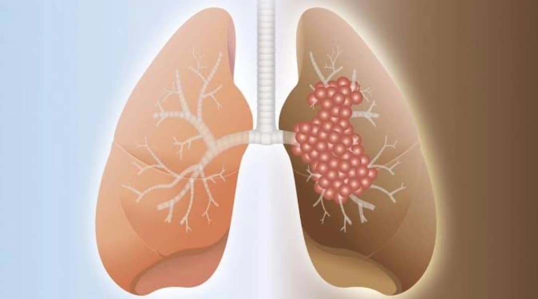
Common early symptoms of lung fibrosis include gradually increasing breathlessness during activity and a persistent dry cough. Some people also develop fatigue, reduced exercise capacity or finger clubbing. Pulmonary fibrosis means that part of the lung tissue has become scarred, making the lungs less flexible and reducing efficient gas exchange. Established fibrosis is usually not fully reversible, but identifying the underlying disease or exposure matters because treatment can sometimes control inflammation, prevent avoidable exposure and slow further progression.
Breathlessness from lung fibrosis often starts quietly.
At first, you may notice it only while climbing stairs, walking uphill or trying to keep pace with other people. Because these changes develop gradually, they are easily blamed on age, weight, a recent infection or lack of fitness.
A persistent dry cough may appear at the same time.
These symptoms are not specific to fibrosis, which is why the diagnosis depends on the whole picture: symptoms, examination, high-resolution CT, lung-function testing, exposure history and, when necessary, additional investigations.
Interstitial lung disease is particularly important in India because its causes and exposures may differ from those reported in Western populations.
In the ILD-India registry of 1,084 newly diagnosed patients from 27 centres, hypersensitivity pneumonitis was the most frequent diagnosis at 47.3%, followed by connective-tissue-disease-associated ILD at 13.9% and idiopathic pulmonary fibrosis at 13.7%.
Those figures describe patients enrolled in that registry and should not be interpreted as the exact prevalence of each type of ILD across the entire Indian population.
What are the early symptoms of lung fibrosis?
Two of the most common symptoms are:
-
Increasing breathlessness during physical activity
-
Persistent dry cough
The important clue is progression.
You may find that an activity you previously managed comfortably gradually becomes more difficult.
Other possible symptoms and signs include:
-
Reduced exercise tolerance
-
Persistent fatigue
-
Faster breathing
-
Loss of appetite or unintentional weight loss in some people
-
Finger clubbing
-
Low oxygen levels, especially during exertion
-
Breathlessness at rest in more advanced disease
Not every person develops all of these features.
Breathlessness on exertion
Early lung fibrosis may first become noticeable when climbing stairs, walking quickly or exercising.
As disease progresses, less strenuous activity may cause breathlessness.
However, shortness of breath has many other causes, including asthma, COPD, heart disease, anaemia and physical deconditioning.
If breathlessness is persistent or getting worse, our guide on when to see a pulmonologist for cough and breathlessness explains when respiratory evaluation is appropriate.
Persistent dry cough
A persistent non-productive cough is common in several fibrotic interstitial lung diseases, particularly idiopathic pulmonary fibrosis.
However, cough alone does not diagnose fibrosis.
Asthma, nasal disease, reflux, medicines, infection and other lung conditions can all cause chronic cough.
Finger clubbing
Clubbing causes the tips of the fingers to become broader and the nails more curved.
It can occur in pulmonary fibrosis, particularly IPF, but it is not present in every patient.
Clubbing can also occur with other lung, heart and gastrointestinal diseases and therefore needs medical assessment rather than being treated as a specific sign of fibrosis.
Fine inspiratory crackles
Doctors sometimes hear fine crackling sounds, particularly over the lower lungs, when listening with a stethoscope.
They are often described as "Velcro-like" crackles.
These sounds can be an important clue to interstitial lung disease but are not diagnostic on their own.
Can lung fibrosis cause symptoms outside the lungs?
Yes, particularly when fibrosis is associated with an autoimmune or connective-tissue disease.
Symptoms worth mentioning to your doctor include:
-
Joint pain or swelling
-
Morning stiffness
-
Fingers changing colour in cold weather
-
Dry eyes or dry mouth
-
Skin tightening
-
Muscle weakness
-
Persistent skin rashes
-
Difficulty swallowing
These clues can point towards conditions such as rheumatoid arthritis, systemic sclerosis, Sjögren disease or inflammatory muscle disease.
Sometimes the lung disease appears before the underlying autoimmune condition has been formally diagnosed.
Is lung fibrosis different from asthma and COPD?
Yes, but symptoms can overlap.
|
Feature |
Fibrotic ILD |
Asthma |
COPD |
|
Breathlessness |
Often progressively worse with activity |
Often variable or episodic |
Usually progressive, particularly with exertion |
|
Cough |
Often dry |
Dry or productive |
Frequently productive |
|
Wheezing |
Less typical but can occur |
Common |
Common |
|
Crackles |
Fine inspiratory crackles may occur |
Usually absent |
May occur for other reasons |
|
Lung-function pattern |
Restriction and/or reduced gas transfer may occur |
Variable airflow obstruction |
Persistent airflow obstruction |
|
Key imaging |
HRCT may show interstitial abnormalities or fibrosis |
Often normal between attacks |
May show emphysema or other changes |
These distinctions are not absolute. For a fuller comparison of the other two conditions, see our guide to asthma vs COPD.
For example, a patient can have both emphysema and pulmonary fibrosis, and lung-function results can occasionally appear relatively preserved despite significant disease.
This is why symptoms alone should not be used to decide whether someone has asthma, COPD or pulmonary fibrosis.
What causes lung fibrosis?
"Pulmonary fibrosis" describes scarring. It does not identify the cause.
Fibrosis can develop as part of several different diseases.
Important groups include:
Hypersensitivity pneumonitis
Hypersensitivity pneumonitis, or HP, is an immune-mediated lung disease caused by sensitisation to inhaled antigens in susceptible people.
Possible exposures include:
-
Birds
-
Feathers
-
Mould
-
Agricultural materials
-
Some occupational organic dusts
-
Contaminated cooling or humidifying systems
The ILD-India registry reported substantial exposure to birds, air coolers, air conditioners and visible mould among patients diagnosed with HP.
However, millions of people have these exposures without developing HP.
Finding an exposure therefore does not prove that it caused an individual's lung disease. The exposure history has to fit with HRCT findings and the rest of the clinical evaluation.
Autoimmune disease
Interstitial lung disease can occur with:
-
Rheumatoid arthritis
-
Systemic sclerosis
-
Sjögren disease
-
Inflammatory myositis
-
Mixed connective-tissue disease
-
Other autoimmune disorders
In some people, the lung problem is detected before other autoimmune symptoms become obvious.
Idiopathic pulmonary fibrosis
Idiopathic pulmonary fibrosis, or IPF, is a progressive fibrotic interstitial lung disease in which no secondary cause such as autoimmune disease, occupational exposure or medication can explain the fibrosis.
It occurs mainly in older adults.
IPF is only one type of pulmonary fibrosis.
Occupational exposures
Long-term exposure to certain dusts can cause fibrotic lung disease.
Examples include:
-
Silica
-
Asbestos
-
Coal dust
-
Other mineral or industrial dusts
A detailed occupational history can therefore be as important as laboratory testing.
Medicines and radiotherapy
Some medicines can cause interstitial lung injury in susceptible people.
Examples include certain:
-
Anti-arrhythmic medicines
-
Antibiotics
-
Chemotherapy drugs
-
Immunomodulatory medicines
Radiotherapy involving the chest can also cause lung injury and fibrosis.
Never stop a prescribed medicine merely because it appears on a list of drugs associated with ILD. Drug-related lung disease requires clinical assessment.
Can pigeons and air coolers cause lung disease?
They can be relevant exposures in hypersensitivity pneumonitis.
Bird proteins and microbial or mould antigens associated with damp environments or inadequately maintained cooling systems can trigger HP in susceptible individuals.
In the ILD-India registry analysis, bird exposure had the strongest association with HP, while air coolers, air conditioners and visible mould were also associated with the diagnosis.
That does not mean every person with pigeons nearby or a desert cooler will develop lung disease.
If HP is suspected, the doctor may ask detailed questions about:
-
Pigeons nesting around the home
-
Pet birds
-
Poultry exposure
-
Feather bedding
-
Damp walls or mould
-
Desert coolers
-
Air-conditioning systems
-
Farming
-
Grain or hay
-
Workplace dusts
The aim is to identify a medically plausible antigen that fits the disease pattern.
What does "fibrotic changes" on a chest X-ray mean?
The phrase "fibrotic changes" on an X-ray does not necessarily mean that you have progressive pulmonary fibrosis.
A localised scar can remain after:
-
Previous tuberculosis
-
Pneumonia
-
Previous inflammation
-
Other old lung injury
This can be very different from a diffuse fibrosing interstitial lung disease affecting larger areas of both lungs.
A chest X-ray also cannot reliably characterise many forms of early interstitial lung disease.
If symptoms or imaging raise concern, the doctor may recommend high-resolution CT.
Can a normal chest X-ray rule out lung fibrosis?
No.
A chest X-ray can miss subtle or early interstitial abnormalities.
When clinical suspicion remains significant, high-resolution computed tomography, or HRCT, provides far more detailed information about the lung tissue.
That does not mean everyone with breathlessness or dry cough needs an HRCT.
The decision depends on clinical examination, symptoms and initial investigations.
How is pulmonary fibrosis diagnosed?
There is no single test that diagnoses every type of pulmonary fibrosis.
Doctors usually combine several pieces of information.
1. Detailed medical and exposure history
This includes:
-
Duration and progression of symptoms
-
Smoking history
-
Bird exposure
-
Mould or dampness
-
Cooling systems
-
Occupational exposures
-
Medicines
-
Radiotherapy
-
Autoimmune symptoms
-
Family history of pulmonary fibrosis
2. High-resolution CT chest
HRCT is central to evaluating suspected fibrotic ILD.
It can show:
-
Reticulation
-
Traction bronchiectasis
-
Honeycombing
-
Ground-glass abnormalities
-
Mosaic attenuation
-
Air trapping
-
Distribution of disease within the lungs
A basal and peripheral pattern with features of usual interstitial pneumonia, or UIP, may support an IPF diagnosis after other potential causes of a UIP pattern have been considered.
Honeycombing alone does not automatically mean IPF.
Similarly, mosaic attenuation and air trapping may support hypersensitivity pneumonitis in the appropriate setting but are not specific enough to diagnose it by themselves.
3. Pulmonary function tests
PFTs help measure how lung disease is affecting respiratory function and are also useful for monitoring change over time.
Forced vital capacity, or FVC, measures how much air can be forcefully exhaled after a full inspiration.
Many fibrotic ILDs produce a restrictive pattern, but normal or near-normal spirometry does not completely rule out early disease.
4. DLCO
The DLCO test measures the ability of gases to move from the air sacs into the bloodstream.
DLCO is commonly reduced in interstitial lung disease and can help assess disease severity and progression.
However, a low DLCO is not specific to fibrosis. Emphysema, pulmonary vascular disease, anaemia and other conditions can also reduce it.
5. Exercise oxygen assessment
A six-minute walk test can assess:
-
Walking distance
-
Symptoms
-
Heart-rate response
-
Oxygen saturation during exercise
Some people maintain normal oxygen levels at rest but desaturate while walking.
6. Blood tests
Blood tests can help look for autoimmune diseases associated with ILD.
The exact panel depends on the patient's symptoms and clinical findings rather than using the same large panel for everyone.
7. Bronchoscopy or lung biopsy
Not every patient needs bronchoscopy or lung biopsy.
Bronchoalveolar lavage may provide useful information in selected cases, including suspected hypersensitivity pneumonitis or infection.
Tissue sampling is considered when the diagnosis remains uncertain and the expected benefit outweighs the procedure risk.
Read our guide to what bronchoscopy involves if your pulmonologist has recommended this test.
Why multidisciplinary review matters
Interstitial lung disease diagnosis can be difficult.
Radiologists, pulmonologists, rheumatologists and pathologists may need to interpret the clinical history, HRCT pattern and laboratory or biopsy findings together.
The ILD-India registry itself found meaningful disagreement between initial diagnoses and expert-panel diagnoses.
For difficult cases, multidisciplinary discussion can substantially improve diagnostic confidence.
Can lung fibrosis be reversed?
It depends on what is meant by "fibrosis" and what disease is causing it.
Established pulmonary fibrosis is generally not fully reversible.
However, many ILDs contain a mixture of fibrosis and potentially treatable inflammation.
Depending on the disease:
-
Removing a relevant antigen may help hypersensitivity pneumonitis stabilise
-
Treating an autoimmune disease can control ongoing inflammatory lung injury
-
Stopping a causative drug may prevent further injury
-
Antifibrotic medicines can slow progression in appropriate patients
Earlier diagnosis is valuable because treatment is generally more effective at preventing additional injury than at restoring lung tissue that has already become permanently scarred.
Is lung fibrosis curable?
There is no single cure that applies to every fibrotic ILD.
Some exposure-related, inflammatory or drug-induced ILDs can improve substantially once the underlying cause is treated.
IPF is a chronic progressive disease for which current drug treatment aims mainly to slow loss of lung function.
Lung transplantation can be considered for selected patients with advanced disease, but transplantation is a major treatment with its own eligibility criteria and risks and should not be described simply as a "cure".
What treatments are available for lung fibrosis?
Treatment depends on the underlying diagnosis.
Hypersensitivity pneumonitis
The first priority is identifying and avoiding a clinically relevant antigen when possible.
Some patients with active inflammatory disease may also require corticosteroids or other immunomodulating medicines.
Treatment of fibrotic HP is more complicated, and not every patient benefits from the same immunosuppressive approach.
If the fibrosis continues to progress despite appropriate management, antifibrotic therapy may be considered in suitable patients.
Connective-tissue-disease-associated ILD
Treatment is tailored to the underlying autoimmune disease.
Medicines may include agents such as:
-
Mycophenolate
-
Cyclophosphamide
-
Rituximab
-
Other disease-specific immunomodulatory therapies
The choice should generally involve both respiratory and rheumatology assessment where appropriate.
Some patients with progressive fibrotic disease may also be candidates for antifibrotic treatment.
Idiopathic pulmonary fibrosis
Established antifibrotic treatments include:
-
Nintedanib
-
Pirfenidone
These medicines do not remove existing scar tissue and patients may not feel an immediate improvement.
Their principal benefit is slowing the rate of lung-function decline.
Treatment requires monitoring for side effects and drug interactions.
Long-term routine immunosuppressive treatment is not used to treat stable IPF in the way it may be used for some autoimmune or inflammatory ILDs.
Progressive pulmonary fibrosis
Progressive pulmonary fibrosis, or PPF, describes progression occurring in an ILD other than IPF despite appropriate management.
Current international guidance defines PPF using a combination of worsening symptoms, physiological progression and radiological progression, after alternative explanations have been considered.
The 2022 ATS/ERS/JRS/ALAT guideline made a conditional recommendation for nintedanib for PPF.
Management of the underlying ILD remains important. "PPF" is a behaviour pattern, not a replacement for identifying the underlying disease.
What is nerandomilast?
Nerandomilast is a newer oral phosphodiesterase-4B inhibitor.
In the 2025 FIBRONEER-IPF trial, nerandomilast produced a smaller decline in FVC over 52 weeks than placebo.
A separate FIBRONEER-ILD trial showed a smaller FVC decline in patients with progressive pulmonary fibrosis.
The US FDA approved nerandomilast, under the brand name Jascayd, for:
-
Adult idiopathic pulmonary fibrosis in October 2025
-
Adult progressive pulmonary fibrosis in December 2025
These approvals are important, but they should not be interpreted as meaning the drug reverses fibrosis.
Indian approval, availability and pricing should be confirmed from current Indian regulatory and prescribing sources before the medicine is presented as a treatment option available locally.
Patients should not obtain or combine antifibrotic medicines without specialist supervision.
Does antifibrotic treatment increase life expectancy?
Antifibrotic treatment has clearly been shown to slow decline in lung function in IPF.
Individual observational studies have also reported differences in outcomes and survival, but it is safer not to promise that a particular medicine will add a specific number of years to an individual patient's life.
Prognosis varies widely depending on:
-
Type of ILD
-
Age
-
Lung function
-
Rate of progression
-
Oxygen requirements
-
Other medical conditions
-
Response to treatment
-
Acute exacerbations
Historical statements that everyone with IPF has only "three to five years" after diagnosis are too simplistic for counselling an individual patient today.
Supportive treatment for pulmonary fibrosis
Treatment is not limited to medication.
Pulmonary rehabilitation
Pulmonary rehabilitation combines supervised exercise, education and breathing strategies.
It can improve exercise capacity, symptoms and day-to-day functioning even though it does not remove fibrosis on the CT scan.
Oxygen therapy
Supplemental oxygen may be prescribed when clinically significant low oxygen levels occur at rest, during activity or during sleep.
The decision should be based on proper oxygen assessment rather than buying oxygen solely because of breathlessness.
Vaccination
Vaccination against relevant respiratory infections is commonly recommended because infections can cause significant deterioration in people with chronic lung disease.
The exact vaccines required depend on age, medical history and current national recommendations.
Smoking cessation
Smoking should be stopped.
That includes cigarettes, bidi and hookah.
Lung transplantation
Patients with progressive advanced fibrotic disease may benefit from early referral to a transplant centre to determine eligibility.
Referral does not mean that transplantation is immediately required. Assessment takes time and is best considered before the disease becomes critically advanced.
When is breathlessness an emergency?
Seek urgent medical attention for:
-
Sudden or rapidly worsening breathlessness
-
Severe breathlessness at rest
-
Blue or grey lips
-
New confusion
-
Fainting
-
Significant chest pain
-
Coughing up substantial blood
-
A new major fall in oxygen saturation, particularly when accompanied by worsening symptoms
-
High fever with worsening breathing
A pulse oximeter can provide useful information, but a single reading should be interpreted alongside symptoms and the person's usual oxygen level.
Someone with known ILD who deteriorates rapidly may have infection, pulmonary embolism, pneumothorax, heart problems, an acute exacerbation of ILD or another complication requiring urgent assessment.
Frequently asked questions
What is usually the first sign of lung fibrosis?
Gradually increasing breathlessness during activity is one of the most common early symptoms.
A persistent dry cough is also common.
Neither symptom is specific to fibrosis, so persistent or worsening symptoms need clinical assessment rather than self-diagnosis.
Is pulmonary fibrosis the same as ILD?
Not exactly.
Interstitial lung disease, or ILD, is the larger group of diseases.
Some ILDs are mainly inflammatory, some are fibrotic, and many contain varying amounts of both inflammation and fibrosis.
Pulmonary fibrosis refers specifically to scarring within the lungs.
For a broader explanation, read our patient guide to interstitial lung diseases.
Is IPF the same as pulmonary fibrosis?
No.
IPF stands for idiopathic pulmonary fibrosis and is one specific type of fibrotic interstitial lung disease.
Many other diseases can also produce pulmonary fibrosis, including autoimmune ILD, hypersensitivity pneumonitis and occupational lung disease.
How long can someone live with pulmonary fibrosis?
There is no single life-expectancy figure for all pulmonary fibrosis.
Different ILDs behave very differently.
Some diseases may remain stable for years, while others progress more quickly.
Even within IPF, progression varies substantially from person to person.
The most useful information for an individual patient comes from the exact diagnosis, serial lung-function tests, HRCT findings, exercise capacity and the rate at which these change over time.
Lung fibrosis ke shuruaati lakshan kya hain?
Lung fibrosis ke common shuruaati lakshan mein chalne, seedhiyan chadhne ya physical activity ke waqt dheere-dheere badhti saans ki takleef aur lagatar sookhi khansi shamil hain.
Ye symptoms sirf fibrosis mein nahi hote. Asthma, heart disease aur doosri lung conditions mein bhi ho sakte hain.
Agar saans ki problem ya khansi lagatar badh rahi ho, to pulmonologist se evaluation karwana chahiye. Zarurat ke hisaab se doctor HRCT aur lung-function tests advise kar sakte hain.
Can pigeons cause pulmonary fibrosis?
Bird exposure can trigger hypersensitivity pneumonitis in susceptible people, and persistent HP can become fibrotic.
But the presence of pigeons alone does not prove that they caused someone's fibrosis.
Diagnosis requires the exposure history to fit with the clinical findings, HRCT pattern and other investigations.
Can an air cooler cause lung fibrosis?
Contaminated cooling systems have been reported as potential antigen sources in hypersensitivity pneumonitis.
Indian registry data found air-cooler exposure commonly among people diagnosed with HP.
However, using a cooler does not automatically mean someone will develop HP or pulmonary fibrosis.
The significance of any suspected exposure must be assessed in the context of the individual's disease.
Diagnose the type, not just the scar
"Pulmonary fibrosis" is a description, not the complete diagnosis.
The important questions are:
-
What caused the fibrosis?
-
Is there active inflammation?
-
Is there an avoidable exposure?
-
Is the disease stable or progressing?
-
Does the patient need immunomodulatory treatment, antifibrotic treatment, supportive therapy or a combination?
Those answers determine treatment far more accurately than the word "fibrosis" on an X-ray report.
Jindal Chest Clinics in Sector 20D, Chandigarh, provides evaluation of interstitial lung disease, pulmonary function testing, DLCO testing, bronchoscopy and specialist review of chest imaging.
Book an appointment or call 0172-4911000 or +91 9779030507.
